Echo Strain: Why It’s So Straining (Pun Intended)
If you’ve been anywhere near an echocardiography lab lately, you’ve probably heard the buzzword: strain imaging. Global longitudinal strain (GLS) is the shiny new tool (that isn't really all that new - it's been around since 2004). Cardiologists can’t get enough of it — and for good reason. It’s sensitive, reproducible, and gives us a better look at subtle myocardial dysfunction than ejection fraction alone.
But let’s be honest: for the sonographer, strain imaging can feel a little… well… straining.
So, What is Strain Anyway?
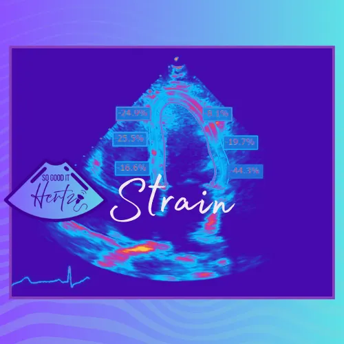
In simple terms, strain measures myocardial deformation — how much the heart muscle fibers shorten, lengthen, or thicken during the cardiac cycle. GLS specifically tracks the longitudinal shortening of the left ventricle, expressed as a percentage. A more negative number (e.g., –20%) = stronger function. A less negative number (e.g., –14%) can be a red flag for early dysfunction, especially in patients receiving chemotherapy or with suspected cardiomyopathy.
Sounds great, right? Until you’re the one actually trying to acquire the images - but it is getting easier and easier with the integration of AI and auto-strain options.
Why It’s Straining for Sonographers
- Frame Rate Drama: You need 40–90 Hz for quality speckle tracking. Too low? Garbage strain curves. Too high? Enjoy trying to get the whole LV in the sector.
- Foreshortening Frustration: If your apical 4-chamber is even slightly off, your bull’s-eye plot looks like a toddler’s art project.
- Patient Cooperation: “Deep breath and hold” quickly turns into deep breath, cough, wiggle, sigh.
- Vendor Variability: Every machine swears it’s the best at strain — but somehow the numbers never quite match from one vendor to the next.
And then there’s the learning curve (almost as straining as the segmental strain curves). Who knew measuring heart muscle deformation could make your own muscles ache?
Professional Tips (So You Don’t Lose Your Mind)
- Be Consistent: Always acquire from standard apical views — A4C, A2C, and A3C — with minimal foreshortening.
- Check Your ECG: Tall, clean R waves make for better tracking. Garbage 💩 in = garbage out.
- Optimize Your Image First: Good endocardial definition is half the battle. Contrast agents can help if your LV walls are playing hide-and-seek - yes there is now contrast strain imaging available (specifically on the Siemens ACUSON Origin)
- Don’t Obsess Over One Curve: Look at the big picture — global strain values matter more than one rogue segment, but you can't ignore the outliers either.
Humor Break
They call it strain because:
- You strain your eyes watching the tracking dots dance across the myocardium.
- You strain your patience when your bull’s-eye looks like abstract art.
- You strain your sanity when the cardiologist says, “Can you just re-do the strain on that patient?” (Again.)
The Takeaway
Echo strain is powerful, valuable, and here to stay. It helps detect dysfunction earlier, track chemo-related cardiotoxicity, and guide patient management in ways ejection fraction alone can’t.
Yes, it can be “straining” for the sonographer — but with good images, careful technique, and a sense of humor, you can master it without pulling a muscle of your own. Because at the end of the day, strain might be tough… but you’re tougher. 💪🫀
-Lara Williams, BS, ACS, RCCS, RDCS, RVT, RDMS, FASE
Don't forget to check out the other platforms below and click that LEARN button up top to check out All About Ultrasound for access to FREE CME!
YouTube: https://www.youtube.com/@SonographersAfterDark
TikTok: https://www.tiktok.com/@sonographersafterdark
Facebook: https://www.facebook.com/groups/sonographersafterdark
Instagram: https://www.instagram.com/sonographersafterdark/
