Hemodialysis Access & Upper Extremity Duplex: A Sonographer’s Playbook
Hemodialysis Access & Upper Extremity Duplex: A Sonographer’s Playbook
Arteriovenous fistulas and grafts are lifelines—your scan can be the difference between a smooth run on the machine and a missed treatment. A crisp, efficient duplex of the upper extremity gives nephrology and vascular teams the roadmap they need to keep access flowing. Here’s a practical, clinic-ready guide you can use today.
What We’re Solving
- Pre-op mapping: Is there adequate inflow artery and a usable, superficial vein for an AVF?
- Post-op checks and maturation: Is flow robust and the vein maturing toward cannulation?
- Troubleshooting: Why is the access alarming, infiltrating, or running at low Qb?
- Post-intervention surveillance: Did angioplasty or thrombectomy fix the real problem?
Pre-Op Mapping Basics
- Patient prep/position: Supine or semi-reclined, arm abducted and supported. Keep the room warm; vasospasm is your enemy.
- Arterial inflow survey: Brachial → radial/ulnar. Record diameter, plaque, and triphasic/biphasic character. Document PSV and any focal accelerations.
- Venous mapping (tourniquet or fist clench): Compressibility, continuity, and diameter at multiple levels (≥2.5–3.0 mm is commonly targeted for native AVF creation). Note depth from skin (shallower is better for cannulation), and mark branches/stenoses.
- Hand perfusion screen: If available, document palmar arch flow (Doppler or plethysmography) when radial-based fistula is considered.
Post-Creation & Maturation Check (AVF/AVG)
- Interrogate the whole circuit: Inflow artery → anastomosis → outflow vein/graft body → central veins (cephalic arch, subclavian) if symptoms suggest.
- Standard measurements:
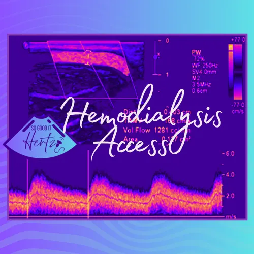
- PSV/EDV with angle correction (≈60°), avoiding the turbulent jet right at the anastomosis for “representative” velocities.
- Volume flow (mL/min): Time-averaged mean velocity × cross-sectional area in a straight, laminar segment (sample multiple sites and average).
- Maturation expectation (the “rule of 6s” for native AVF):
- Diameter ≥ 6 mm, Depth ≤ 6 mm, Flow ≥ 600 mL/min by ~6 weeks (institutional criteria vary).
- Grafts typically mature faster; depth is usually ideal, so focus on flow and stenosis detection.
Red Flags You Can’t Miss
- Juxta-anastomotic stenosis (common in AVF): Focal high PSV with a velocity ratio ≥2:1 suggests ≈50% diameter reduction; ≥3:1 suggests ≈75%. Expect post-stenotic turbulence and drop in volume flow.
- Cephalic arch stenosis (brachiocephalic AVF): Look for focal acceleration and waveform change; correlates well with recirculation and high venous pressures on dialysis.
- Outflow vein problems: Long-segment narrowing, kinks at transposition sites, or cannulation-site aneurysms/pseudoaneurysms with mural thrombus.
- Inflow insufficiency: Damped waveforms proximal to the anastomosis, small arterial caliber, or diffuse disease lowering access flow.
- Thrombosis/near-occlusion: Echogenic intraluminal material, no color fill, absent/markedly reduced spectral signal.
- Steal syndrome: Reduced digital arterial signals, symptomatic hand coolness/pain; compare pre- and post-fistula waveforms and consider reactive maneuvers (gently compress outflow and watch digital improvement).
- High-output concern: Very high access flows (>1500–2000 mL/min) plus systemic symptoms may warrant correlation with cardiac function.
Technique Pearls
- Angle and gate care: Keep your angle at ~60°, gate centered, and sample a clean laminar segment for flow. Slow your sweep speed to stabilize envelope tracing for accurate TAMV.
- Measure more than once: Average 2–3 volume-flow sites; flows fluctuate with hydration and blood pressure.
- Scan lightly: Especially over fresh AVFs—minimal probe pressure preserves true caliber and avoids spasm.
- Map cannulation zones: Note straight, superficial segments ≥6–8 cm for two-needle use; annotate depth so staff can select needle length wisely.
- Central veins when indicated: If arm/face swelling, high recirculation, or persistent alarms, screen the cephalic arch/subclavian; escalate to dedicated central venous imaging if suspicion remains.
Common Access Types (Know Their Quirks)
- Radiocephalic (wrist): Great longevity, but more juxta-anastomotic stenosis and maturation failures in small vessels.
- Brachiocephalic (antecubital): Watch the cephalic arch; high turbulence and stenosis are frequent.
- Brachiobasilic transposition: Deep course pre-transposition; after surgery, track the entire elevated segment for kinks or focal narrowings.
- AV grafts (PTFE): Expect higher baseline velocities and more pseudoaneurysms at cannulation sites; assess both arterial and venous anastomoses closely.
What to Put in Your Final Impression
- Access type and laterality (e.g., left brachiocephalic AVF).
- Global flow (mL/min) and whether it meets maturation/clinical targets.
- Hemodynamically significant lesions with location (e.g., 2.7:1 PSV ratio at juxta-anastomotic segment, flow drop distal).
- Complications (aneurysm/pseudoaneurysm, thrombus, perigraft fluid/hematoma).
- Actionable recommendation aligned with your lab’s criteria (e.g., “Findings consistent with ≥50% outflow stenosis—correlate for angioplasty,” or “Flow and caliber meet maturation criteria—suitable cannulation segment from mid-forearm to antecubital fossa, depth 3–4 mm”).
Fast Checklist Before You Sign Off
- Inflow artery PSV/EDV and plaque note
- Anastomosis view and representative velocities
- Outflow caliber, depth, and volume flow (≥2 sites)
- Velocity ratios across any focal lesions
- Cannulation map and depth
- Complications documented with cine or clips
How Your Scan Helps the Team
- Dialysis staff can choose better needle sites (fewer infiltrations, better Qb).
- Surgeons/interventionalists can target the exact lesion, reducing contrast and procedure time.
- Patients keep their lifeline—fewer missed treatments and fewer catheters.
Dialysis access is dynamic—vessels remodel, stenoses evolve, and flows change with the patient’s day. With a disciplined duplex protocol, clear numbers, and a laser-focused impression, you become the guardian of that circuit. Keep it patent, keep it painless, and keep it flowing.
Stay laminar, trust your ratios, and may your waveforms be so good it hertz.
For more details and SDMS CME CreditUpper Extremity Duplex and Hemodialysis Access - Check out All About Ultrasound's CME Course
-Lara Williams, BS, ACS, RCCS, RDCS, RVT, RDMS, FASE
Don't forget to check out the other platforms below and click that LEARN button up top to check out All About Ultrasound for access to FREE CME!
YouTube: https://www.youtube.com/@SonographersAfterDark
TikTok: https://www.tiktok.com/@sonographersafterdark
Facebook: https://www.facebook.com/groups/sonographersafterdark
Instagram: https://www.instagram.com/sonographersafterdark/