Fetal Echocardiography: Mastering the 3-Vessel View
Fetal Echocardiography: Mastering the 3-Vessel View
Fetal echocardiography is a powerful tool for detecting congenital heart disease (CHD) before a baby even takes their first breath. Among the essential cardiac planes, the 3-Vessel View (3VV) is one of the most important — and sometimes one of the trickiest — to master. When obtained correctly, it provides a quick, reliable assessment of the fetal great vessels and their relationship in the upper mediastinum.
Why the 3-Vessel View Matters
The 3VV allows you to evaluate the size, arrangement, and flow direction of the:
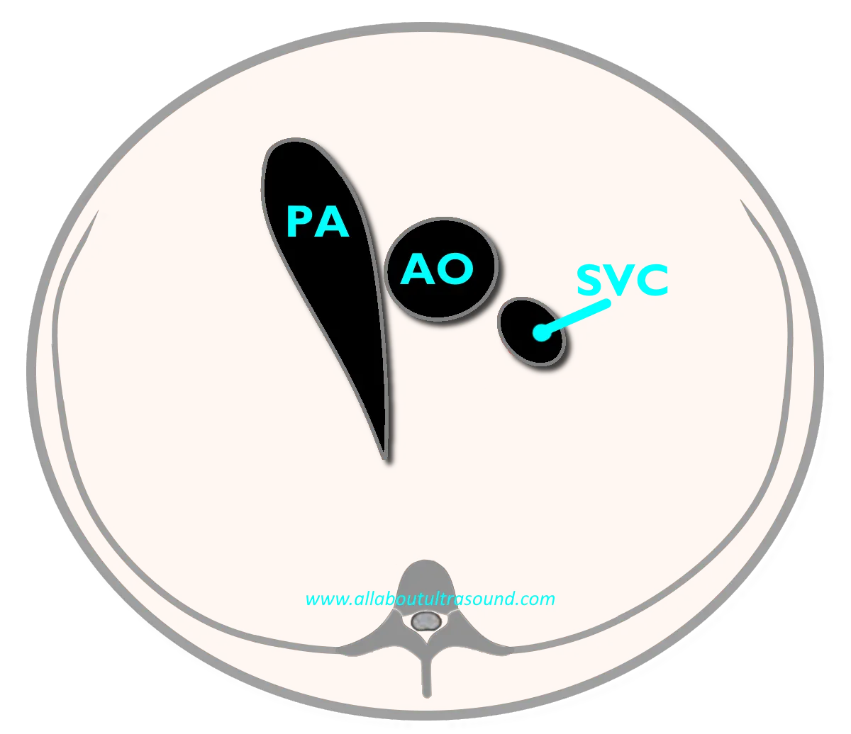
- Main Pulmonary Artery (MPA) - The largest and most anterior vessel.
- Ascending Aorta (AO) - Located centrally.
- Superior Vena Cava (SVC) - The smallest and most posterior vessel.
When normal, these vessels line up from left to right in a decreasing size pattern: PA (largest) → AO (medium) → SVC (smallest). Their arrangement and relative size can give early clues to congenital abnormalities like transposition of the great arteries, truncus arteriosus, right or left outflow tract obstruction, or even arch anomalies.
Think of it as the traffic report of fetal circulation — you want to know which lanes are open, which are narrowed, and whether someone is going the wrong way.
How to Obtain the 3-Vessel View
- Start with the Four-Chamber View: From here, sweep cranially (toward the fetal head).
- Angle Slightly Upward: As you move cephalad, the left ventricular outflow tract transitions to the short axis of the great vessels.
- Identify the Landmarks:
- The pulmonary artery should be anterior and to the left.
- The aorta sits just to the right of the pulmonary artery.
- The SVC is posterior and rightward.
- Use Color Doppler: Flow direction confirms anatomy — look for laminar forward flow in all three vessels.
- Freeze and Optimize: Adjust depth, zoom, and gain so the three vessels sit crisp and clear across the top of your screen.
Tips and Tricks for Success
- Patience with Fetal Position: Sometimes the fetus is curled, spine-up, or just uncooperative (future toddlers in training). Give the patient time, ask them to walk, or rescan later if needed.
- Adjust Your Angle: Small changes in transducer tilt can mean the difference between seeing a beautiful 3VV and staring at rib shadows.
- Mind the Scale: Color Doppler can be your best friend, but set the Nyquist limit appropriately to avoid aliasing.
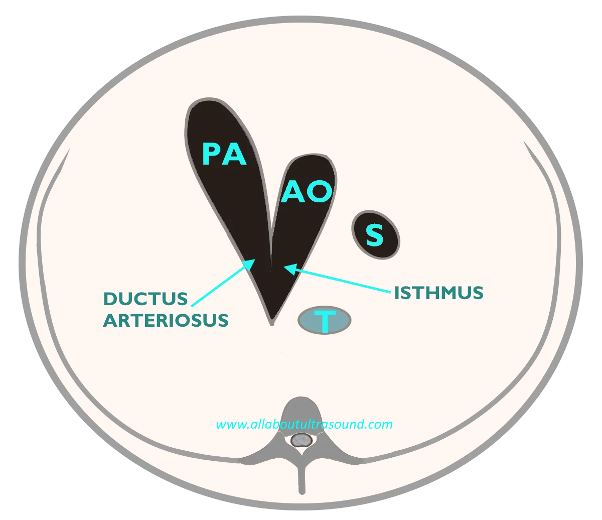
- Don’t Forget the 3VT (3-Vessel Trachea) View: A quick sweep cranially from the 3VV shows the great vessels curving into the aortic and ductal arches — crucial for ruling out arch abnormalities.
Humor Break
If you’ve ever spent 10 minutes trying to coax a fetus into the perfect position for the 3VV, you know the drill. One minute the vessels are right there, the next minute the baby flips and you’re staring at a spine that looks like it’s laughing at you. It’s like trying to take a passport photo of a toddler — just when you think you’ve got it, they move.
The Takeaway🎯
The 3-Vessel View is a cornerstone of fetal echocardiography, offering a window into the great arteries that can reveal critical CHD early. With practice, patience, and solid technique, sonographers can consistently obtain this view and provide physicians with the diagnostic confidence they need.
✨Remember: PA → AO → SVC, left to right, largest to smallest. Get that pattern down, and you’ll never forget it.
-Lara Williams, BS, ACS, RCCS, RDCS, RVT, RDMS, FASE
Don't forget to check out the other platforms below and click that LEARN button up top to check out All About Ultrasound for access to FREE CME!
YouTube: https://www.youtube.com/@SonographersAfterDark
TikTok: https://www.tiktok.com/@sonographersafterdark
Facebook: https://www.facebook.com/groups/sonographersafterdark